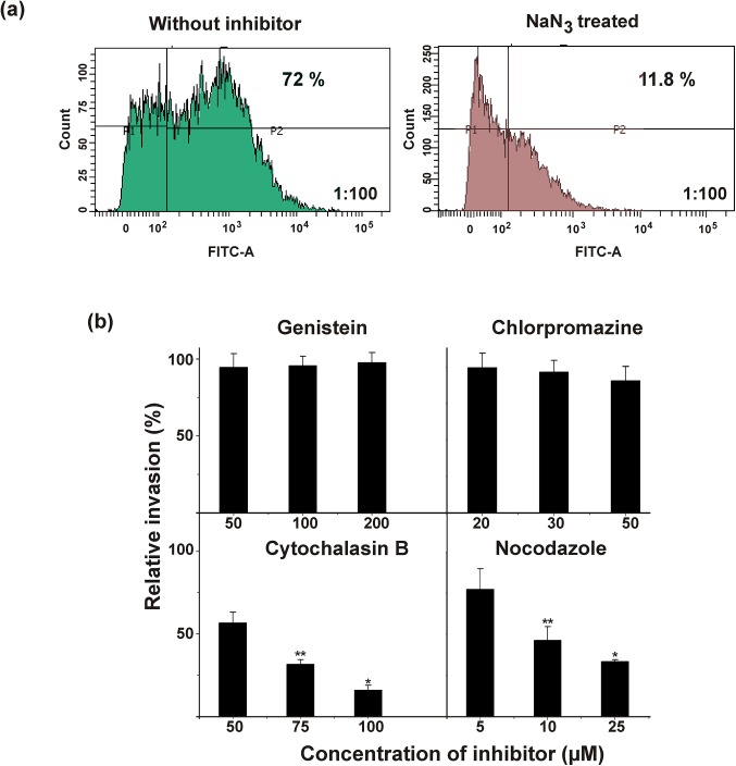
Fig 2. Energy dependent endocytic uptake of S. agalactiae by H9C2 cells.
(a) Quantification of endocytic uptake of fluorescently‒labeled live S. agalactiae (AO-stained) by H9C2 cells in the presence (green) or absence (brown) of sodium azide by flow cytometry. P1-unstained population of H9C2 cells, P2- population of H9C2 cells internalized with fluorescently‒labelled S. agalactiae. (b) Determination of intracellular relative viable CFU of S. agalactiae recovered from H9C2 cells in the presence of different endocytic inhibitors such as genistein, chlorpromazine, cytochalasin B and nocodazole. The results were expressed in relative percentage compared with the experimental control. Statistically significant differences are indicated by an asterisk (* p<0.05 or **p<0.01).

