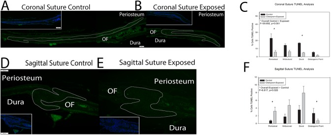
Fig 3. Histological markers of apoptosis in control and citalopram exposed mouse cranial sutures.

A & B. Coronal sutures from control (A) and citalopram exposed (B) 15 day old C57BL6 mice stained for Terminal deoxynucleotidyl transferase dUTP nick end labeling (TUNEL) activity (green). Periosteum, dura and osteogenic front (OF) are as marked, and the sutural area has been outlined. Inset lower magnification of TUNEL staining with a 4',6-diamidino-2-phenylindole (DAPI) nuclear counterstain (blue). Note the highly cellularized (blue stained) area of the suture abutting the less cellularized (darker) area of the osteogenic front. C. Quantification of percent TUNEL positive staining as compared to DAPI stain in coronal sutures. Significant decreases in TUNEL activity were noted due to citalopram exposure at the periosteal and dural areas of the sutures. Midsutral space and osteogenic fronts did not demonstrate significantly different staining between unexposed and citalopram exposed sutures. D & E. Sagittal sutures from unexposed (control) (D) and citalopram exposed (E) 15 day old C57BL6 mice stained for TUNEL activity (green). Inset lower magnification of TUNEL staining with a DAPI nuclear counterstain (blue) F. Quantification of percent TUNEL positive staining as compared to DAPI stain. A significant increase in TUNEL staining was noted in the periosteal area while a significant decrease in staining was noted at the osteogenic front of citalopram exposed sagittal sutures.
