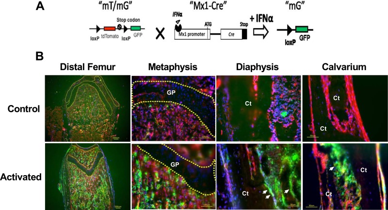
Fig 1. Characterization of Mx1-Cre in the long bones and calvariae in vivo.
(A) A schematic diagram showing that mT/mG is crossed to Mx1-Cre and that IFNα can activate Cre expression to delete the tdTomato (red fluorescent) cassette and initiate GFP (green fluorescent, “mG”) expression. (B) Mx1-Cre;mT/mG mice at the age of 5 weeks were injected intraperitoneally with PBS (control) or pI-pC (activated). The distal femurs and calvariae were collected for cryosection after three days. The nuclei were stained with DAPI (blue). The arrows indicate bone surface GFP expression in the pI-pC-treated bones. The dotted lines circle the cartilage regions in the distal femur. GP, growth plate cartilage; Ct, cortical bone.

