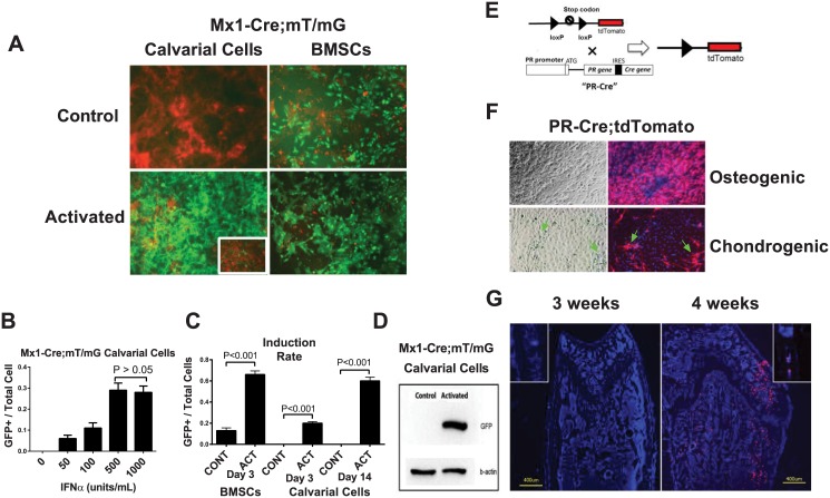
Fig 2. Comparison of BMSCs and calvarial cells in terms of Mx1-Cre activation and PR expression in vitro.
(A) BMSCs or calvarial cells were obtained from Mx1-Cre;mT/mG double transgenic mice and treated with IFNα (500 units/mL) for three days. Fluorescent images were taken three days (BMSCs) or 14 days (Calvarial cells) after the IFNα treatment. The image of calvarial cells three days post IFNα treatment is shown in the insert. The levels of Cre activation were quantitated by GFP (green) expression. (B) A quantitative histogram showing the induction rates (the GFP+ cells versus the total cells) of the BMSCs and calvarial cells shown in Figure A. CONT, control; ACT, activated. (C) Western blotting was performed to detect GFP protein in the total cell lysates from the calvarial cells with (activated) or without (control) IFNα treatment on day 3. β-actin was used as an internal control. (D) Mx1-Cre;mT/mG calvarial cells were treated with different concentrations of IFNα for three days. The ratios of GFP+ cells to total cells under each IFNα concentration were quantified (A). A PR-Cre;tdTomato schematic diagram showing that the Cre gene is inserted downstream of the endogenous PR gene after an internal ribosome entry site (IRES) sequence such that Cre is expressed simultaneously with PR. When Cre is expressed (together with PR), it recombines loxp sites to remove the stop codon before the tdTomato cassette and activates tdTomato (red fluorescence) expression. (E) A diagram of PR-Cre; tdTomato. The tdTomato expression can be activated following the upstream stop codon removal by Cre, which is expressed simultaneously with endogenous PR gene. So that PR+ cells will express tdTomato (red fluorescent). (F) PR-Cre;tdTomato calvarial cells were differentiated into osteoblasts with osteogenic media or chondrocytes with chondrogenic media for 14 days. The majority of the cells cultured in the osteogenic medium were red. However, when the cells were cultured in the chondrogenic medium, only a small percentage of the fibroblast-like cells (arrows) turned red. The nuclei were stained with DAPI (blue). (G) Distal femurs or calvariae (inserts) from 3-week-old or 4-week-old PR-Cre;tdTomato mice were sectioned for fluorescent microscopy. The nuclei were stained with DAPI (blue). The red fluorescence indicated PR promoter activity.

