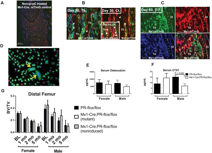
Fig 4. Skeletal phenotypes of Mx1-Cre-driven PR inactivation in vivo.
(A) A distal femur from non-pI-PC treated Mx1-Cre;mT/mG mouse. (B—C) Mx1-Cre;mT/mG mice were injected with pI-pC intraperitoneally to induce Cre at one month of age and then sacrificed one (B) or two (C) months later. The distal femurs (D.F.) were collected and sectioned to observe the GFP (green) and tdTomato (red) fluorescence. Green indicates the Mx1+ cells that expressed Cre, and red indicates the Cre-negative cells. The nuclei were stained with DAPI (blue). The femoral trabecular bones from the Col1a1-CreERT2;mT/mG mice that received 4 days of tamoxifen injections were used as a positive control and are shown in the insert in (B). The white arrows indicate the green osteocytes that were observed in the trabecular bone in the Col1a1-mT/mG mice but were absent in the trabecular or cortical bone of the Mx1-mT/mG mice (the white arrowheads indicate the GFP-/tdTomato+ osteocytes). There were no green osteocytes observed in the Mx1-mT/mG bones on either day 30 or 60. (D) Bone marrow cells were collected from 1-month-old Mx1-Cre;mT/mG mice. Some multinuclear cells turned green indicating Mx1-Cre activation in these cells (yellow arrows). (E) Serum osteocalcin and (F) serum CTX1 levels were measured by ELISA two months post pI-pC injection (n = 8/group). (G) Five-week-old male and female Mx1-Cre;PR-flox/flox or PR-flox/flox (control) mice were injected with pI-pC intraperitoneally to induce Cre activity. Baseline microCT scans (BL) of the distal femurs were performed on five-week-old mice without receiving the pI-pC injection. The animals were sacrificed at one, two or five months post-pI-pC injection, and the hind limbs were collected for microCT analysis (n = 8/group).

