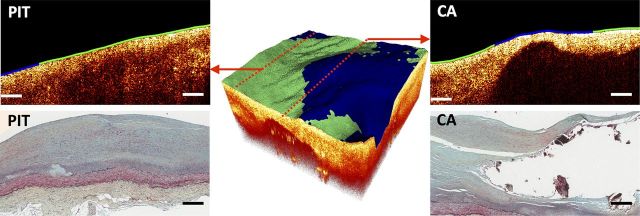
Figure 3.

Sample multimodal OCT–FLIM volume of a CA with a mixed luminal biochemical composition. The colour-coded lumen indicates the superficial plaque composition based on FLIM: HL as red, HC as green, and LCL as blue. An OCT B-scan (left-top) taken from the edge of the volume indicated a PIT with a HC (green) superficial composition, confirmed by histopathology (left-bottom). Another OCT B-scan (right-top) taken from the middle of the volume showed a calcified necrotic core covered by a fibrous cap of mixed LCL (blue, middle) and HC (green, sides) composition, also confirmed by histopathology (right-bottom). All scale bars represent 200 µm.
