
Figure 2.
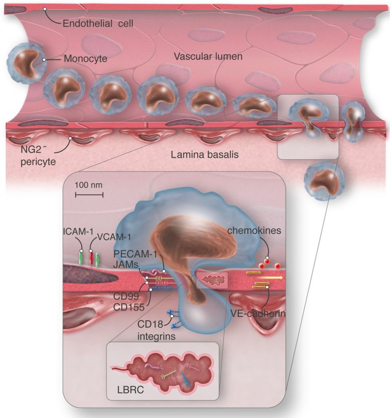
Schematic view of paracellular monocyte transmigration. ICAM-1, intercellular adhesion molecule-1; PECAM-1, platelet endothelial cell adhesion molecule; CD99, cluster of differentiation 99; CD155, cluster of differentiation 155; VE-cadherin, vascular endothelial-cadherin; CD18 integrins, cluster of differentiation 18, beta subunit of integrins LFA-1 and Mac-1; JAMs, junctional adhesion molecules; LBRC, lateral border recycling compartment; NG2−, neuron-glial-2 negative. Vascular endothelial cell, red; monocyte, light blue; pericytes, red-brown. Monocyte extravasation into the site of inflammation requires transendothelial migration and penetration of the lamina basalis NG2− pericytes. Monocytes transmigrate at sites of low matrix protein density. Monocyte engagement of endothelial cell surface molecules activates targeted recycling of the LBCR and thus enlarges the transmigration gap. The LBRC (brown) contains PECAM-1, CD99, CD155, and JAM-A, but not VE-cadherin, and supplies the transmigrating monocyte with additional functional molecules. VE-cadherin stabilizes junctional integrity in steady-state and transiently abandons site of transmigration under inflammatory conditions.