Fig. 3.
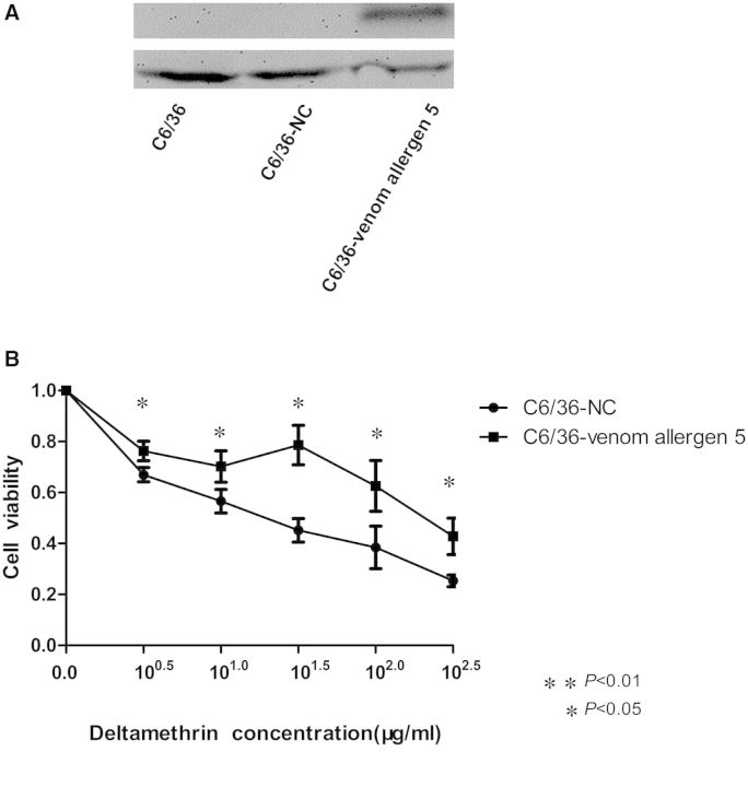
In vitro validation by plasmid transfection and cytotoxicity assay. (A) Results of western blot analysis of the protein expression of venom allergen 5 in the C6/36, C6/36-NC, and C6/36-venom allergen 5 cells. β-actin was used as the internal control. The C6/36-venom allergen 5 cells expressed the corresponding protein of ∼32 kDa. The experiment was repeated three times. (B) Cytotoxicity assay using a CCK-8 kit. The x-axis shows the five concentrations of deltamethrin. The y-axis gives cell viability (as a proportion of the total cells relative to a 0 μg/ml concentration). The viability of the C6/36-venom allergen 5 cells was significantly higher than the C6/36-NC cells with each of the five deltamethrin concentrations. Figures show the mean ± SD of three independent experiments. *P < 0.05, **P < 0.01 compared with the C6/36-NC cells.

