
Figure 6.
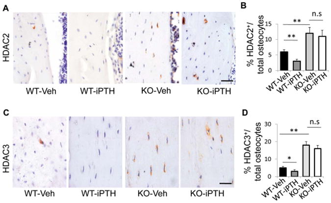
Inhibitory effect of PTH on HDAC 2 and 3 protein expression in osteocytes is abolished by LRP6 deficiency. (A) Representative images of immunohistochemical staining with an antibody to HDAC2 (brown) in 3-month-old male WT and KO mice treated with vehicle or PTH1–34 daily at 80 mg/kg bw, 5 days a week for 4 weeks. Scale bars: 50μm. (B) Quantification of HDAC2+ osteocytes out of total osteocytes; n = 6; *P < 0.05, **P < 0.01. (C) Representative images of immunohistochemical staining with an antibody to HDAC3 (brown) in 3-month-old male WT and KO mice treated with vehicle or PTH1–34 daily at 80 mg/kg bw, 5 days a week for 4 weeks. Scale bars: 50 μm. (D) Quantification of HDAC3+ osteocytes out of total osteocytes. A total of three femur sections from each mouse, and six mice per treatment group were analyzed. *P < 0.01.