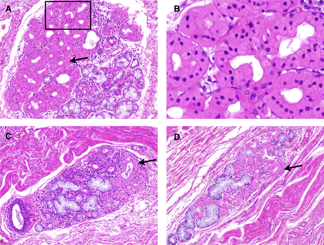
Figure 2.
ESMGs containing oncocytic acini from controls: Oncocytes appear densely eosinophilic with a centrally located nucleus. A. 100× view of an ESMG containing 70% oncocytic acini, present on the left (arrows). Mucinous acini, characterized by their pale mucosa are present right side of this ESMG. B. 200× view of oncocytes from Panel A. C. 40× view of ESMG that contains 25% oncocytic acini. D. 40× view of ESMG containing 62% oncocytic acini.

