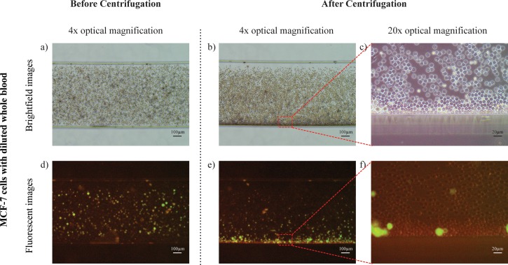
FIG. 5.
Cells separation in the centrifugal microfluidic device. MCF-7 cells mixed with diluted whole blood were loaded into a new microfluidic chip. Representative images of MCF-7 cells with diluted whole blood under brightfield microscopy within the separation channel (a) before centrifugation and (b) after centrifugation. (c) Representative image under 20× optical magnification at the bottom of the separation channel indicating MCF-7 cells migration. (d)–(f) Corresponding images under fluorescence microscopy indicating the separation of the MCF-7 cells.

