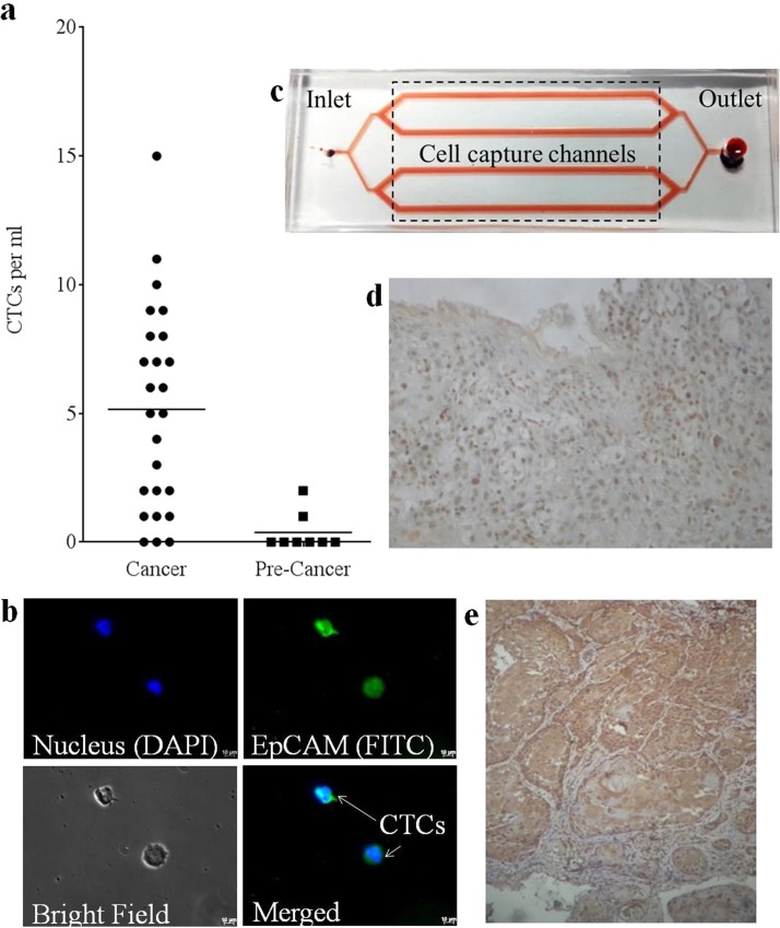
FIG. 6.
CTC capture and characterization from blood samples of head and neck cancer patients: (a) Quantification of CTCs captured from blood samples of patients with cancer and pre-cancerous lesions. (b) Representative fluorescence microscopy images of CTCs captured from blood sample of head and neck cancer patients. (c) Layout of the microfluidic device immobilized with EpCAM LNA aptamer used for capture of CTCs from cancer samples. Photomicrograph showing the cytoplasmic and nuclear localization of EpCAM ICD in (d) gingival squamous cell carcinoma (20× magnification) and (e) tongue squamous cell carcinoma (10× magnification).

