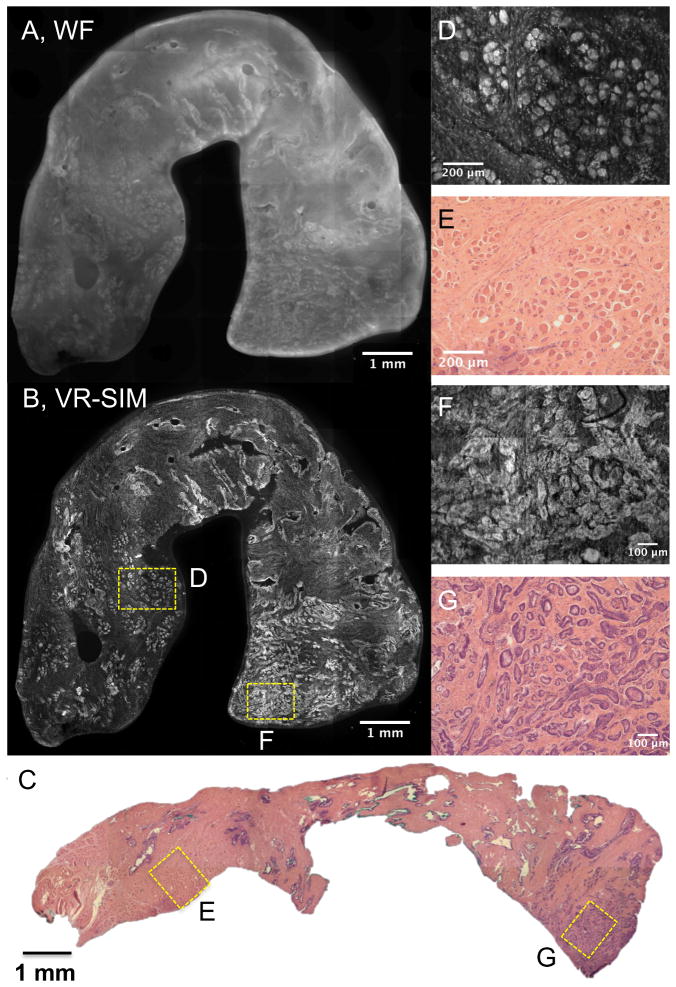
Figure 1.
VR-SIM images and subsequent H&E slide images of Biopsy A1 confirmed as malignant. A) Wide-field (i.e., without SIM) image of the entire biopsy, B) VR-SIM mosaic image of the entire biopsy, comprising 205.5 megapixels. C) Digital image of the corresponding H&E section. D) VR-SIM and E) H&E zoom images of the regions of interest marked by the correspondingly labeled boxes in B and C, depicting an area of normal skeletal muscle and fibrous stroma. F) VR-SIM and G) H&E zoom images of the regions of interest marked by the correspondingly labeled boxes in B and C, depicting an area of malignant glands.

