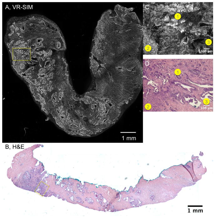
Figure 2.
VR-SIM images and subsequent H&E slide images of biopsy A19 confirmed as malignant. A) VR-SIM mosaic image of the entire biopsy, comprising 205.5 megapixels, B) Digital image of the corresponding H&E section. Zooms of the dashed yellow boxes in A and B are shown in C and D, respectively. Corresponding areas of interest between the VR-SIM image and the H&E image are denoted by numbered circles (1 = Gleason grade 3 cancer, 2 = Gleason grade 4 cancer, and 3 = benign glands).

