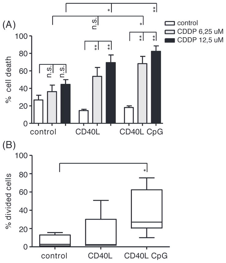
Figure 1.

CDDP treatment of resting and dividing CLL cells. (A) Apoptosis after 48 h of treatment with CDDP as indicated in PB derived CLL cells stimulated with CD40L ± GpC for 4 days, assessed by MitoTracker staining (n = 6); mean ± standard error of mean (SEM); *p < 0.05, **p < 0.01 (Mann–Whitney test). (B) Percentage of CLL cells proliferating upon stimulation with CD40L and CpG for 4 days (n = 6) as indicated (and as described in “Materials and methods” section); mean, whiskers min to max; *p < 0.05 (Mann–Whitney test).
