Figure 5.
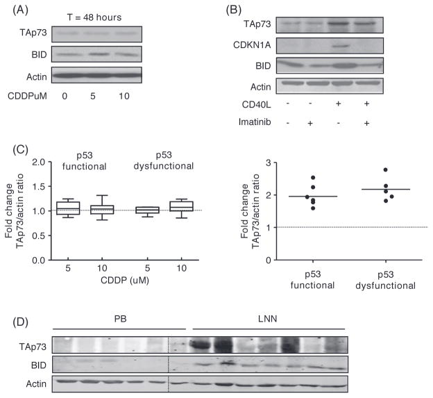
Differential expression of TAp73 in PB versus LN derived CLL cells. (A) Protein levels of TAp73 and BID assessed by Western blot in CLL cells with dysfunctional p53 after CDDP treatment for 48 h as indicated. Actin was used as loading control. (B) Protein levels of TAp73, CDKN1A and BID in CLL cells cocultured with CD40-ligand expressing fibroblasts for 24 h with or without imatinib. Cells were washed and lysed after an additional 24 h of culture. Actin was used as loading control. (C) Fold induction in TAp73/actin ratio upon CDDP treatment for 48 h as indicated in p53 functional (n = 7) and p53 dysfunctional (n = 8) CLL (left panel) or after CD40-ligand stimulation in p53 functional (n = 6) and p53 dysfunctional (n = 5) CLL (right panel). (D) Protein levels of TAp73 and BID in peripheral blood (PB) and lymph node (LNN) derived CLL cells. Actin was used as a loading control.

