
Fig. 2.
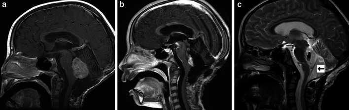
Preoperative and postoperative MRI images from patient 2. a Preoperative sagittal T1-weighted MRI with gadolinium demonstrating recurrent, enhancing tumor filling fourth ventricle. b Postoperative sagittal T1-weighted MRI with gadolinium demonstrating subtotal resection of tumor. A small amount of tumor adherent to the floor of the fourth ventricle was purposefully left behind. c Postoperative T2-weighted sagittal MRI demonstrating catheter position within the fourth ventricle. The catheter, as shown by the arrow, is positioned in a trajectory such that injury to the brainstem and cerebellum are avoided and all catheter holes are within the fourth ventricle