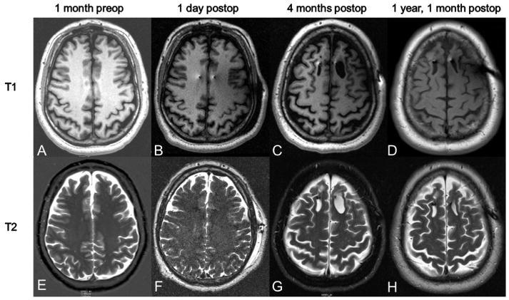
FIG. 1.
T1 and T2 post imaging sequences. Imaging was obtained 1 month preoperatively and then at several time points postoperatively. Postoperative time points included 1 day, 4 months, and 1 year, 1 month. No edema or abnormalities are seen on the 1 day postop scans. The cystic lesion was first noted on the 4 month postop scan and is visible in both T1 and T2 sequences. The lesion is noted to be stable at 1 year and 1 month postoperatively, with slight involution of the left frontal lesion.

