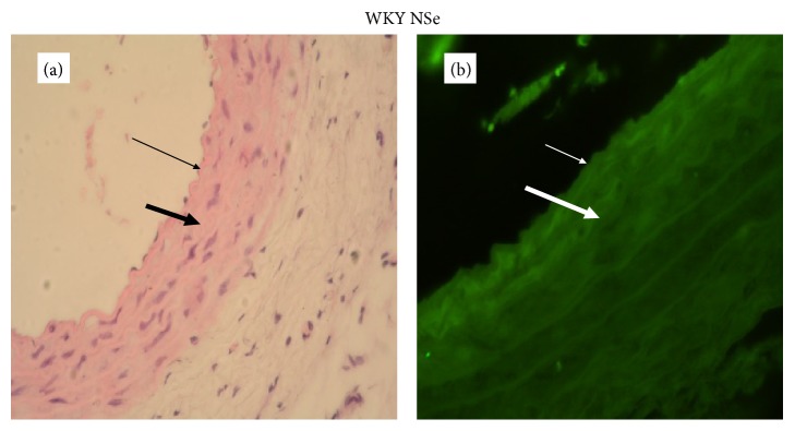
Figure 4.

A representative photomicrograph of aortic wall of WKY on adequate selenium content diet (thin arrow: endothelium; thick arrow: media). (a) Histology, intact endothelium and normal thickness of aortic wall (magnification ×400). (b) Expression of GPx-1, immunofluorescent lighting with moderate intensity (magnification ×400).
