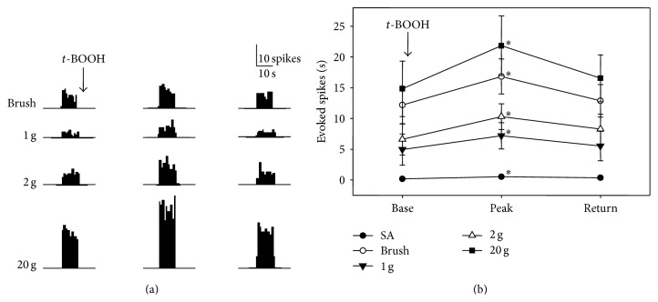
Figure 2.
Effect of t-BOOH on WDR neuron activities in the spinal dorsal horn of normal rats. (a) The neurons responded well to a variety of mechanical stimuli applied to the receptive field before administration of t-BOOH (left column). After local application of 1 μmol of t-BOOH in 10 μL to the spinal cord without washout, the discharge rates started to increase, peaked by 60.0 (±14.1) min (center column), and then returned to the baseline levels by 122.1 (±32.1) min (right column). Stimulus duration is indicated by horizontal bars at the bottom. (b) Evoked responses of WDR neurons (n = 7) to various stimuli before and after t-BOOH application. Asterisks indicate values that are significantly different from the baseline values as determined by a one-way repeated-measures analysis of variance followed by the Duncan test (P < 0.05). The data are mean with standard errors of the mean.

