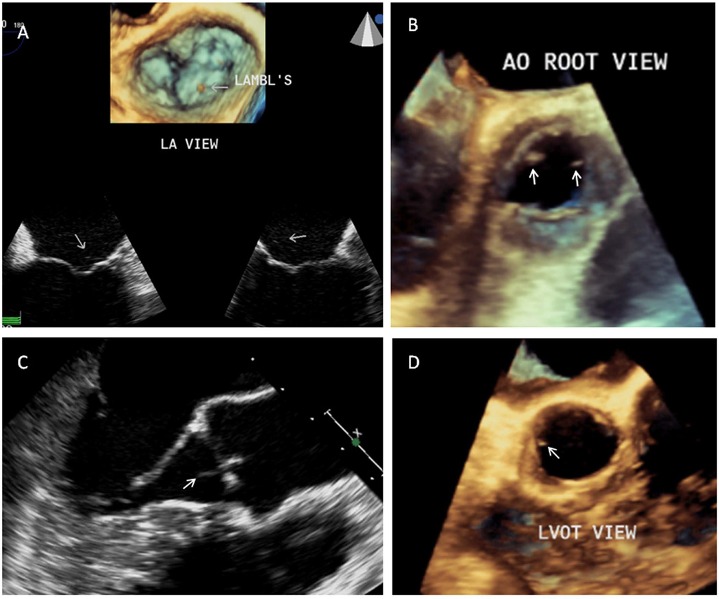
Figure 1. Lambl’s excrescences of the mitral and aortic valves.
A. Two-dimensional (2D) TEE 4 and 2-chamber views of the mitral valve (bottom images) in a 21 year-old healthy female demonstrate a long, thin, and hypermobile Lambl’s excrescence located at the coaptation point and atrial side of normal mitral leaflets (Figure 1A video clip). Corresponding three-dimensional (3D) TEE atrial view of the mitral valve (top image) shows a Lambl’s excrescence at the coaptation point of the middle anterior and posterior mitral scallops (Figure 1A video clip). B. This aortic root 3D-TEE view of the aortic valve in a 44 year-old female with SLE demonstrates a Lambl’s excrescence (right arrow) on the tip and ventricular side of the left coronary cusp and a Libman-Sacks vegetation (left arrow) located on the tip and ventricular side of a thickened non-coronary cusp (Figure 1B video clip). C,D. This longitudinal 2-D TEE view (C) of the aortic valve in a 34 year-old female with SLE demonstrates a thin, elongated, and mobile Lambl’s excrescence at the coaptation point of the left and right coronary cusps prolapsing into the ventricular outflow tract during diastole (arrow) (Figure 1C video clip). Corresponding left ventricular outflow tract (LVOT) 3D-TEE systolic view (D) demonstrates a Lambl’s excrescence located on the ventricular side and tip of the left coronary cusp (Figure 1D video clip).

