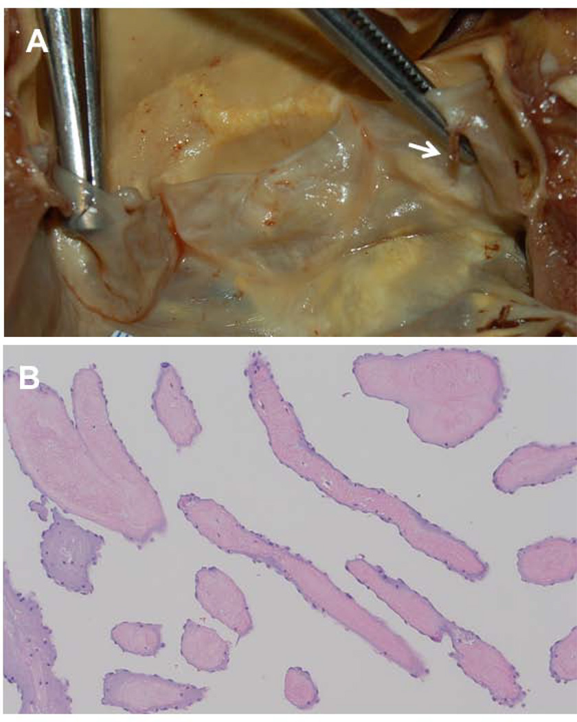
Figure 2. Histology of Lambl’s Excrescences.
A. Gross anatomic view of a mildly sclerotic aortic valve with a thin (1 mm) and elongated (6 mm) Lambl’s excrescence (arrow) at the coaptation point and ventricular side of the left coronary cusp. B. Histology of longitudinal, cross-sectional, and oblique cuts of the Lambl’s excrescence demonstrate a core of fibroelastic, hypocellular, and avascular connective tissue covered by a single layer of endothelial cells.

