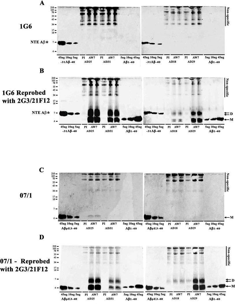
Fig. 4. Aβ in AD-TBS extracts contains little pE3 Aβ and no detectible N-terminally extended Aβ species.
TBS extracts from the same four AD brains shown in Figures 2 and 3 were IP’d with AW7 or pre-immune serum (PI) and WB’d with 1G6 (A) or mAb 07/1 (C). Recombinantly produced −31Aβ-40 (5, 10 and 45 ng) included on gels in panel A were readily detected, whereas no 1G6 immunoreactive species were detected in AD-TBS samples. Importantly, when the same blots were reprobed with a combination of 2G3 and 21F12 (B), abundant ~4 and ~7 kDa species were detected. AβpE3-40 (5, 10 and 45 ng) standards were readily detected with 07/1. A mAb 07/1-immunoreactive band that co-migrated with synthetic AβpE3-40 was detected in one of the 4 AD brains examined. When the same blots were reprobed with a combination of 2G3 and 21F12 (D), abundant ~4 and ~7 kDa species were detected. AD brain numbers and the use of PI or AW7 for IP is indicated below each lane. Molecular weight markers are shown on the left. M denotes monomer and D, dimer. * indicates N-terminally extended (NTE) −31Aβ-40 standard.

