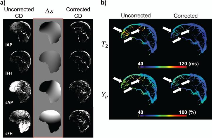
Figure 2.
The effect of eddy-current correction. (a) A set of representative uncorrected and corrected CD images, as well as the Δε hyperplane calculated for the correction algorithm. Shown are the eTE=0ms images for all four TRU-PC scans (sensitized to different flow directions and blood velocities). lAP: large vessel in Anterior-Posterior, lFH: large vessel in Foot-Head, sAP: small vessel in Anterior-Posterior, sFH: small vessel in Foot-Head. (b) The uncorrected and corrected T2 maps (in ms), and respective Yv maps (in %). The eddy-current correction modifies the T2 and Yv (white arrows).

