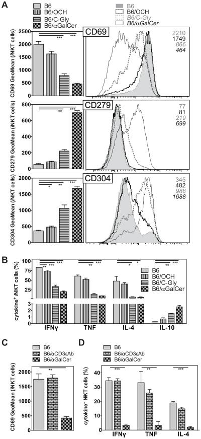
Figure 2. iNKT cell hypo-responsiveness does not solely depend on strong TCR - mediated activation.
(A, B) C57BL/6 (B6) mice were either left untreated or injected i.v. with 4μg of OCH, C-Gly or αGalCer as indicated. One month later mice were injected i.v. with 1μg αGalCer, and 90 min later expression surface markers (A) and the production of indicated cytokines (B) by splenic iNKT cells was analyzed. For (A) representative data (right panel) and summary graphs (left panel) are shown. The utilized gating strategy for iNKT cells is depicted in Supplemental Fig. 1. (C, D) C57BL/6 (B6) mice were either left untreated or i.v. injected with 4μg αGalCer or 1μg of αCD3ε (145.2C11) antibodies as indicated. One month later mice were injected i.v. with 1μg αGalCer, and 90 min later expression of CD69 (D) and of indicated cytokines (E) by splenic iNKT cells was analyzed. Statistically significant differences of treated groups versus the control group are indicated. Representative data from one of two independent experiments are shown.

