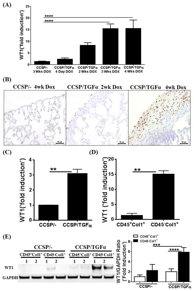
Figure 1. Wilms’ tumor 1 (WT1)-positive cells accumulate in the subpleural fibrotic lung lesions of TGFα mice.
(A) WT1 transcripts were quantified by RT-PCR in the lungs of CCSP/− or CCSP/TGFα transgenic mice fed doxycyclin (Dox) food for 4 days or 2, 3, and 4 wks (n=6). (B) Immunostaining of lung sections with anti-WT1 antibodies shows progressive accumulation of WT1-expressing cells in the subpleura of CCSP/TGF-α mice on Dox for 2 and 4 wks compared to CCSP/− mice on Dox for 4 wks. Images are representative of n=4 per group. Scale bar, 50 μm. (C) WT1 transcripts were quantified by RT-PCR in the total RNA of lung mesenchymal cell cultures from CCSP/− and CCSP/TGFα mice fed on Dox food for 4 wks. (D) The total lung mesenchymal cells were separated into fibrocytes (CD45+Col1+) and resident mesenchymal cells (CD45−Col1+) using ant-CD45 magnetic beads from lung mesenchymal-cell cultures of CCSP/− or CCSP/TGF-α mice on Dox for 4 wks, and WT1 transcripts were quantified by RT-PCR. (E) The lysates of fibrocytes (CD45+Col1+) and resident mesenchymal cells (CD45−Col1+) from CCSP/− or CCSP/TGF-α mice on Dox for 4 wks were blotted with anti-WT1 antibodies and quantified using the Phosphor Imager software, and values were normalized with GAPDH control. One-way ANOVA with Sidak’s multiple comparisons test was used to measure significant differences between groups. **P<0.005, ***P<0.0005, ****P<0.0001 Results are representative of two or more independent experiments.

