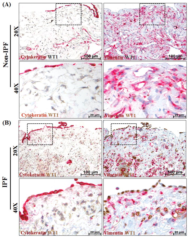
Figure 2. Wilms’ tumor 1 (WT1)-positive cells accumulate in the subpleural fibrotic lesions of lungs in patients with idiopathic pulmonary fibrosis (IPF).
Mesothelial and mesenchymal cells are the major lung cell types that express WT1 in the subpleural fibrotic lesions of the human IPF lung. Serial lung sections from (A) non-IPF (n=4) and (B) IPF (n=6) patients were co-immunostained with antibodies against either cytokeratin (red, indicates mesothelial cells) and WT1 (brown) or vimentin (red, indicates mesenchymal cells) and WT1 (brown). All images were collected at 20X (scale bars, 100 μm) and 40X (scale bars, 20 μm) magnification. The dashed boxes in the 20X images indicate the areas highlighted in the 40X images.

