
Figure 3. Fibrocytes do not transform into Wilms’ tumor 1 (WT1)-positive cells in the subpleural fibrotic lung lesions of TGFα Mice.
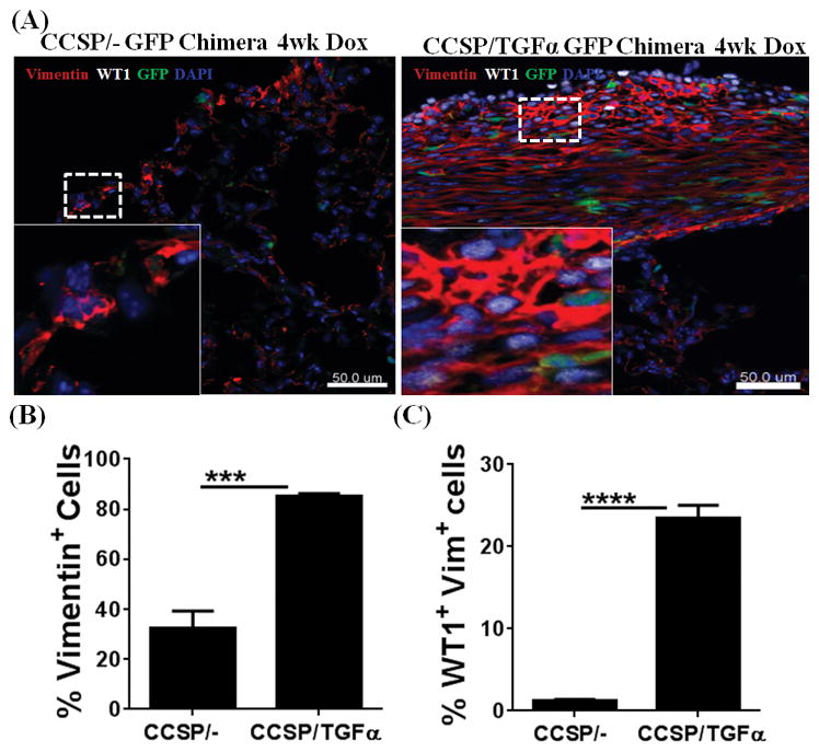
GFP-chimera mice were generated by transplanting 3×10⁶ bone marrow (BM) cells from EGFP-transgenic mice into lethally-irradiated (11.75Gy) CCSP/− and CCSP/TGFα recipients. BM cells were harvested from the tibia and femur of the EGFP donor mice by flushing the bones with Dulbecco’s PBS under aseptic conditions and washing by centrifugation (5 min at 1,000 x g, 4°C). (A) Lung sections from CCSP/− GFP and CCSP/TGFα GFP chimera mice fed doxycyclin (Dox)-treated food for 4 wks were stained with antibodies for WT1 (white) and vimentin (red). The dashed box indicates the enlarged area. Images are representative of n=5 per group. Scale bar, 50 μm. (B) Quantification of the total vimentin-positive cells in the subpleural regions of CCSP/− GFP chimera and CCSP/TGFα chimeric mice using Imaris version 7.2.3 (n=5). (C) Quantification of the total WT1-positive cells from the subpleural regions of CCSP/− GFP chimera and CCSP/TGFα chimeric mice (n=5). Data shown are means ± SEM. Unpaired Student’s t-test was performed to measure the significance. ***P<0.0005, ****P<0.0001