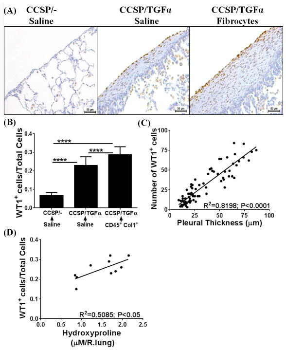
Figure 5. Adoptive cell transfer of fibrocytes augments accumulation of Wilms’ tumor 1 (WT1)-positive cells in the subpleura of fibrotic lung lesions during TGFα-induced pulmonary fibrosis.
CCSP/− and CCSP/TGFα mice were fed with doxycyclin (Dox)-treated food for 14 days and fibrocytes (0.7×10⁶ cells/mice) or saline was infused intravenously via the tail vein. After fibrocyte infusion, mice were continued on Dox for another 7 days and lungs were harvested for analysis. (A) Immunostaining of lung sections with anti-WT1 antibodies. Images are representative of n=4–6 per group. Scale bar, 50 μm. (B) The number of WT1-positive cells per total cells in the lung pleura/sub-pleura was calculated by counting WT1-positive cells and total cells in pleura/sub-pleural regions from five representative images per animal from CCSP/− and CCSP/TGF-α mice (n=4–6/group). Data shown are means + SEM. Statistical significance between groups was measured using One-way ANOVA with Sidak’s multiple comparison test. ****P<0.0001 (C) The correlation was calculated between the number of WT1-positive cells and subpleural thickness using Pearson correlation coefficient with linear regression analysis (r2=0.8198; P<0.0001; n=4–6 per group). (D) The correlation was calculated between the number of WT1-positive cells and hydroxyproline levels of the total lung using Pearson correlation coefficient with linear regression analysis (r2=0.6076; P<0.005; n=4–6 per group).

