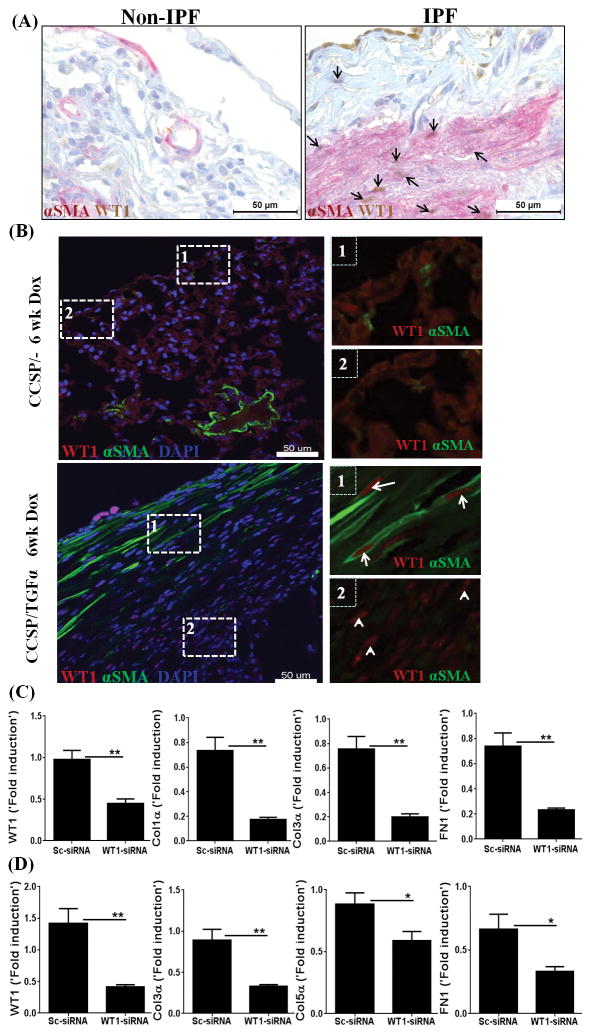
Figure 8. Wilms’ tumor 1 (WT1) is expressed by myofibroblasts and is a critical regulator of extracellular matrix (ECM) gene expression.
(A) Lung sections of non-IPF and IPF patients were co-immunostained with antibodies against αSMA (red) and WT1 (brown). Images are representative of n=4 per group. Scale bar, 50 μm. (B) Immunofluorescence staining for WT1 and αSMA on lung sections of CCSP/− and CCSP/TGFα mice on Dox for 6 wks. The dashed box with the number one is used to indicate the subpleural area with cells positive for both WT1 and αSMA (WT1-positive myofibroblasts). The dashed box with the number two is used to indicate the subpleural area with the cells positive for WT1, but limited or no staining for αSMA. Images are representative of n=4 per group. Scale bar, 50 μm. (C) Primary lung-resident mesenchymal cells were isolated from the subpleural lung mesenchymal cell cultures of human IPF by negative selection using anti-CD45 magnetic beads and transfected with either control or WT1 siRNA for 72 h. The transcripts for WT1 and ECM genes, including Col1α, Col3α1, and FN1, are shown as the fold induced by normalizing to 18s rRNA control. Data shown are mean + SEM values (n=2). Statistical significance between groups was measured using an unpaired Student’s t-test. *P<0.05, **P<0.005 (D) Primary lung-resident mesenchymal cells were isolated from subpleural lung mesenchymal-cell cultures of CCSP/TGFα mice on Dox 4 wks by negative selection using anti-CD45 magnetic beads and transfected with either control or WT1 siRNA for 72 h. The transcripts for WT1 and ECM genes, including Col3α, Col5α, and FN1, are shown as the fold induced by normalizing to a hypoxanthine guanine phosphoribosyl transferase (HPRT) control. Data shown are mean + SEM values (n=4/group). Statistical significance between groups was measured using an unpaired Student’s t-test. *P<0.05, **P<0.005

