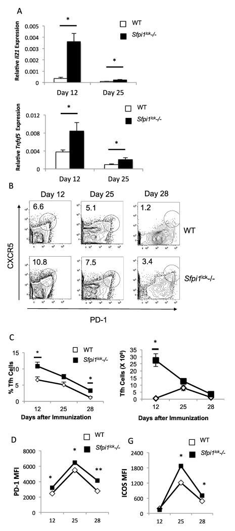
Figure 3.
Sfpi1lck−/− mice have increased Tfh cells after immunization with MOG35-55. WT and Sfpi1lck−/− mice were immunized with MOG35-55 and sacrificed 12, 25, and 28 days after initial immunization. A, Spleens from immunized mice were harvested from wildtype and Sfpi1lck−/− mice for mRNA analysis. mRNA levels of the indicated genes 12 and 25 days after immunization are shown. B, Splenocytes from immunized mice were stained for Tfh markers and analyzed by flow cytometry. C, Percent of Tfh and number of Tfh cells in wild type and Sfpi1lck−/− mice 12, 25 and 28 days after immunization are shown. D, PD-1 and ICOS expression by WT and Sfpi1lck−/− Tfh cells were measured by flow cytometry. Data are representative of 2-3 experiments with 3-6 mice per group (A-D). Statistical significance was determined with a two-tailed t test, *, p<0.05; **, p<0.005.

