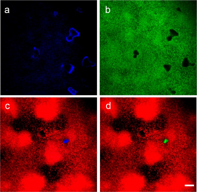
FIG 1.
Imaging of mineral deposits in P. aeruginosa biofilms by confocal laser reflection. Calcium carbonate minerals and biofilm morphology were imaged simultaneously in PAO1-gfp (a and b) and PAO1-mCherry (c and d) biofilms. (a, c, and d) Minerals imaged by laser reflection appear blue (a and c), and those imaged by calcein staining appear green (d). (b, c, and d) Biomass appears green (PAO1-gfp) (b) and red (PAO1-mCherry) (c and d). (c and d) Minerals imaged by laser reflection and calcein staining present identical morphologies. Scale bar = 20 μm.

