Figure 5.

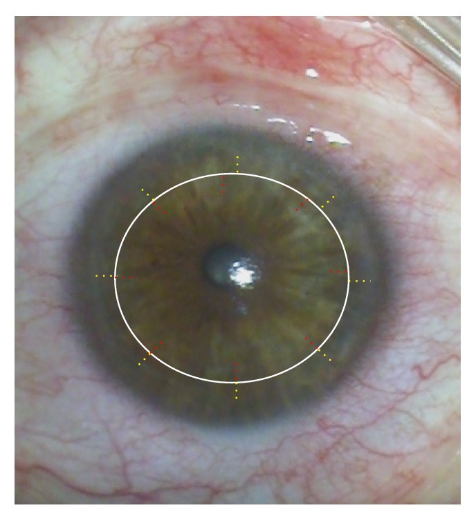
(Hypothetical diagram) different locations of radial incisions in donor and recipient (for better visualization, the red markings are in the donor and the yellow in recipient) after femtosecond laser trephination in keratoconus.

(Hypothetical diagram) different locations of radial incisions in donor and recipient (for better visualization, the red markings are in the donor and the yellow in recipient) after femtosecond laser trephination in keratoconus.