Figure 7.

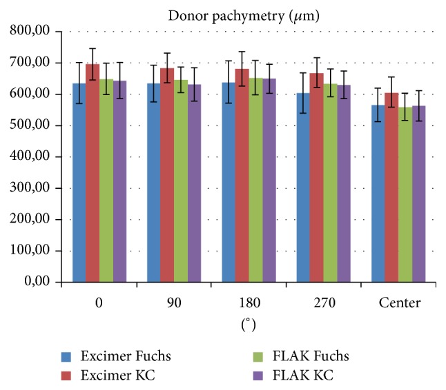
Distribution of pachymetry values for all study groups measured manually with ultrasound pachymetry at the center and in 4 midperipheral points at 0°, 90°, 180°, and 270° (KC = keratoconus, Fuchs = Fuchs endothelial dystrophy).

Distribution of pachymetry values for all study groups measured manually with ultrasound pachymetry at the center and in 4 midperipheral points at 0°, 90°, 180°, and 270° (KC = keratoconus, Fuchs = Fuchs endothelial dystrophy).