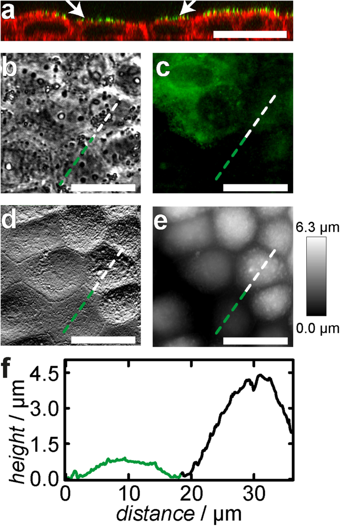
Figure 6. Topography of MDCK II cells after ezrin knock-down via siRNA.
(a) Confocal fluorescence micrograph of the xz plane of a confluent cell layer. The plasma membrane is stained with PKH67 Green Fluorescent Cell Linker, F-actin is labeled with Alexa Fluor 546-phalloidin. Cells without ezrin (arrows) were found to be reduced in height compared with not successfully transfected cells. (b) Phase contrast image. (c) Corresponding fluorescence micrograph showing the ezrin distribution. Ezrin is stained with ezrin mouse IgG primary and Alexa Fluor 488 labeled goat anti-mouse IgG secondary antibody. Some cells (green) were not successfully transfected with siRNA and serve as a control. (d) AFM deflection and height image (e) of the same spot shown in (b,c). (f) Height profile along the green/white dotted line. Cells lacking ezrin are substantially flattened. (Scale bar: 20 μm).

