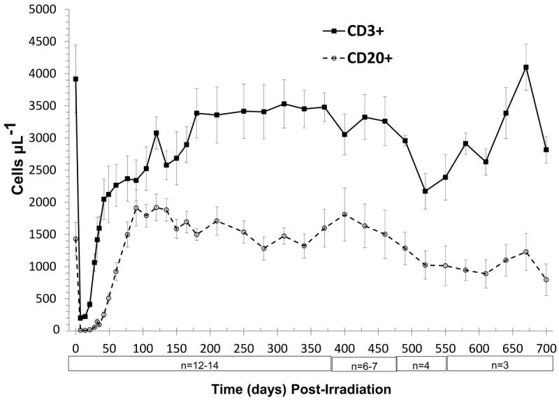
Figure 3.

Time course of peripheral T- and B-lymphocyte loss and recovery in peripheral blood of rhesus macaques following 6.00 Gy total body x-irradiation (mean values ± standard error). Samples were obtained on selected days (n = 12-14 between days 0 to 370, n = 6-7 between days 385 to 460, n = 3-4 between days 475 to 700) following TBI. Whole blood was stained with fluorescently tagged antiCD3, antiCD20 antibodies, red blood cells were lysed, then T- and B-lymphocytes were identified using a flow cytometer, quantified and presented as cells μL-1.
