Fig. 4.
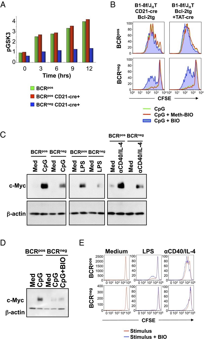
GSK3β activity prevents the proliferation of CpG-treated BCRneg B cells. (A) Splenic B cells from B1-8f/JHT, bcl-2tg or B1-8f/JHT, CD21-Cre, bcl-2tg mice were stimulated with CpG for the indicated times and stained for CD19 and IgM and intracellular phospho-GSK3α/β (S21/9). BCRpos (green) cells were gated from B1-8f/JHT, bcl-2tg cells, whereas Cre+ BCRpos (red) or Cre+ BCRneg (blue) B cells were gated from B1-8f/JHT, CD21-Cre, bcl-2tg cells. Normalized mean fluorescence interval values for phospho-GSK3α/β (S21/9) are shown. Data represent three biological replicates. (B) B cells from B1-8f/JHT, CD21-Cre, bcl-2tg mice or TAT-Cre-transduced B cells from B1-8f/JHT, bcl-2tg mice were CFSE-labeled and stimulated for 3 d with CpG alone (green) or with Meth-BIO (inactive control inhibitor, red) or BIO (GSK3 inhibitor, blue). Cells were gated on ToPro3−, CD19+, and IgM+ or IgM−. Data are representative of three biological replicates. (C) Splenic B cells from B1-8f/+, bcl-2tg mice were TAT-Cre-transduced and cultured for 3 d. Purified BCRpos and BCRneg B cells were placed in culture overnight and stimulated with CpG, LPS, or anti-CD40/IL-4 for 6 h. Equal amounts of protein from whole-cell lysates were used for Western blotting with the indicated antibodies. Data are representative of three biological replicates. (D) Sorted BCRpos and BCRneg B cells from B1-8f/+, CD21-Cre, bcl-2tg mice were cultured in medium, CpG, or CpG and BIO for 6 h. Lysates were analyzed by Western blot for c-Myc and β-actin levels. Data are representative of three biological replicates. (E) Splenic BCRpos and BCRneg B cells from B1-8f/+, CD21-Cre, bcl-2tg mice were sorted, labeled with CFSE, and cultured in media or with LPS or anti-CD40/IL-4 alone (red) or with BIO (blue). Data are representative of three biological replicates.

