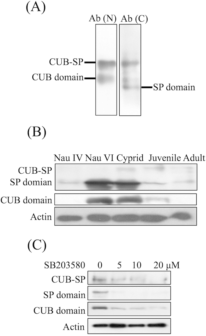
Figure 3. Characterization of CUB-Serine protease expression.

(A) Two bands corresponding to 58 and 33 kDa were detected using an antibody against the CUB domain. Two bands corresponding to 58 and 25 kDa were detected using an antibody against the serine protease domain. Ab(N): antibody raised against the N-terminus of the CUB-serine protease (CUB domain). Ab(C): antibody raised against the C-terminus of the CUB-serine protease (serine protease domain). (B) Western blot showing that the CUB-serine protease was highly expressed in nauplius VI and the cyprid stages. Three bands, including the intact protein, CUB domain and serine protease domain of the CUB-serine protease, were visualized. The signals of the isolated CUB and serine protease domains were much stronger than that of the intact protein. Nau: nauplius. (C) Western blot showing that 5, 10 and 20 μM of SB203580 decreased the level of the intact protein as well as the isolated CUB and serine protease domains of the CUB-serine protease.
