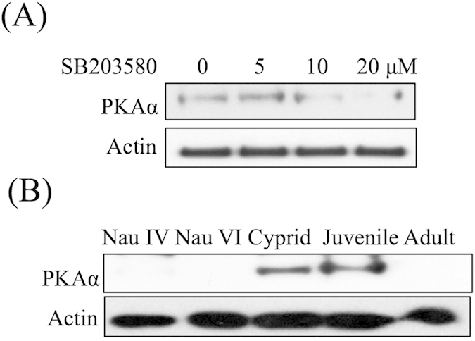
Figure 6. Characterization of PKAα expression.

(A) Western blot showing that the level of PKAα decreased in response to 10 and 20 μM SB203580. (B) PKAα was highly expressed during the cyprid and juvenile stages.

(A) Western blot showing that the level of PKAα decreased in response to 10 and 20 μM SB203580. (B) PKAα was highly expressed during the cyprid and juvenile stages.