
Fig. 2.
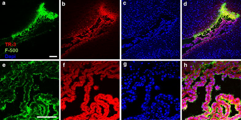
Penetration of TR-3 in the choroid plexus after striatum injection. Horizontal section −3 mm from the bregma. a–d Overview of the lateral ventricle. F-500 accumulated towards the ependymal layer (a) while TR-3 appeared to cross the ependymal more easily (b). Cell nuclei stained with DAPI (c). A merged picture is shown in panel d. e–g Detail of the choroid plexus. F-500 remains confined to the surface of the choroid plexus (e) while TR-3 clearly penetrated the choroid plexus (f). Cell nuclei stained with DAPI (g). Merged picture (h). Scale bar 100 µm