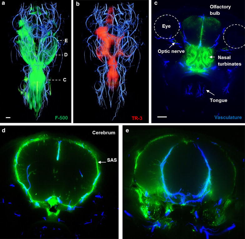
Fig. 5.

Three dimensional reconstruction of the vasculature with co-localized distribution of tracers in a mouse head after injection in the cisterna magna. a F-500 was found in the subarachnoid space and along the penetrating arteries of the brain. b TR-3 was observed in the same compartment, but less confined and also diffusely present in the brain parenchyma. This was particularly evident around the large arteries of the circle of Willis. c Outside the brain, a strong signal was found in the nasal turbinates and along the optic nerves. Maximum intensity projections of 50 images at the indicated levels of the vasculature and F-500 are shown in panels C–E. Panels a, b: dorsal view; c–e: coronal view. Scale bar 1 mm
