
Fig. 1.
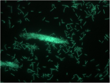
Micrograph of Geobacillus thermoglucosidasius C56-YS93 cells showing individual cells and clumps of cells. Cells were grown in TSB plus 0.4 % glucose for 18 h. at 70 °C. A 1.0 ml aliquot was removed, centrifuged, re-suspended in 0.2 ml of sterile water, and stained using a 50 μM solution of SYTO® 9 fluorescent stain in sterile water (Molecular Probes). Dark field fluorescence microscopy was performed using a Nikon Eclipse TE2000-S epifluorescence microscope at 2000× magnification using a high-pressure Hg light source and a 500 nm emission filter