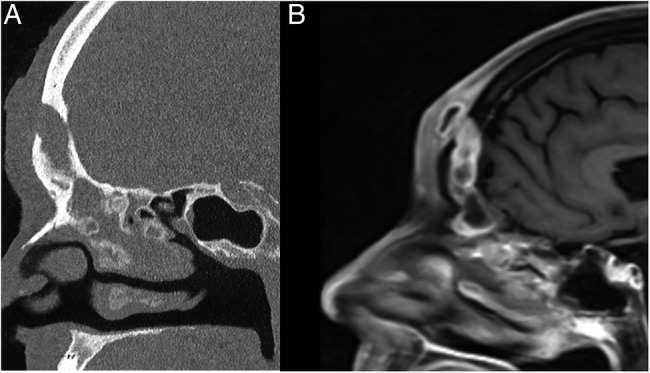
Figure 2.

(A) Sagittal CT of the paranasal sinuses demonstrating an opacified frontal sinus, erosion of the anterior wall of the frontal sinus and contiguous abscess of the soft tissues of the forehead. (B) Sagittal T1-weighted MRI with contrast of the paranasal sinuses showing forehead soft tissue abscess with enhancement in continuity with opacified frontal sinus.
