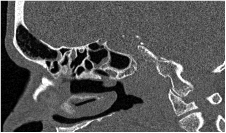
Figure 3.

Post-treatment sagittal CT scan demonstrating a well-pneumatised frontal sinus; the other paranasal sinuses are also healthy with no disease recurrence. The imaging improvement can lag behind clinical response, though in this case, a sufficient time window has elapsed between treatment and repeat CT scan.
