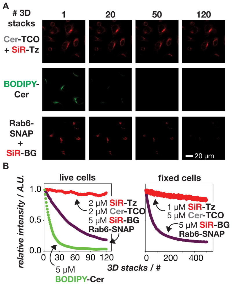
Figure 5.
Golgi labeled with the lipid Cer-SiR is extremely stable to prolonged illumination using spinning disk confocal microscopy. a) Images show cells labeled with Cer-SiR (Cer-TCO+SiR-Tz), BODIPY-Cer, or the protein Rab6-SNAP labeled with SiR (Rab6-SNAP+SiR-BG) after acquisition of 1–120 3D image stacks (22 images/stack). b) Plot of the relative, average per-cell intensity of cells labeled with different lipid and protein probes as a function of the number of acquired 3D stacks.

