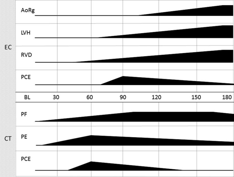
Figure 11.
Making the connection: Time of onset, progression, and severity of lung and cardiac tissue injuries.
The figure represents organ-specific injury initiated at time of radiation and progression to time of onset. Multimodal imaging occurs at 30 day intervals and impacts our ability to correctly identify the duration of the latent period. Subject-based trigger-to-treat medical management also impacts the severity and progression of organ injury. Given these variables the data base suggested certain trends of tissue damage to both organs. Pulmonary fibrosis/pneumonitis (PF) and pleural effusion (PE) first appear at 30 DPI. PF increased throughout study without resolution at 180 DPI. PE spiked in early time points and persisted in some NHP for the study duration. Pericardial effusion (PCE) viewed by computed tomography is observed from 60 DPI to 120 DPI. Cardiac damage appeared later. Aortic regurgitation (AoRg) first appeared at 120 DPI and increased until study conclusion. Left ventricle hypertrophy (LVH) first appeared at 90 DPI and progressed to the end of study. Right ventricle dilation (RVD) first appeared at 60 DPI and worsened throughout study duration. PCE first appeared at 90 DPI and diminished in volume throughout the study as seen by echocardiography (EC).Pulmonary-DEARE appeared to be more sensitive to acute radiation-induced injury than cardiac-DEARE relative to time of onset.

