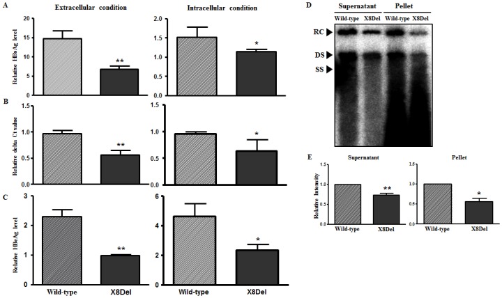
Fig 4. Comparison of the HBsAg secretion and virion formation capacities between the HBV full-length genome constructs pHBV1.2X-Wild and pHBV1.2X-X8Del in HuH-7.5 cells.

After the transient transfection of pHBV-1.2x-Wild type and pHBV1.2X-X8Del into HuH-7.5 cells for 2 days, the levels of HBsAg (A) and HBeAg (C) from the supernatant and the RLB-lysed pellet were measured using the HBsAg and HBeAg ELISA kits. The virion levels in the supernatant and pellet after the transfection of both plasmids were compared using real-time quantitative PCR (B) and Southern blotting analysis (D). The relative intensity of DNA from the Southern blotting was measured using ImageJ software (NIH, USA) (E).
