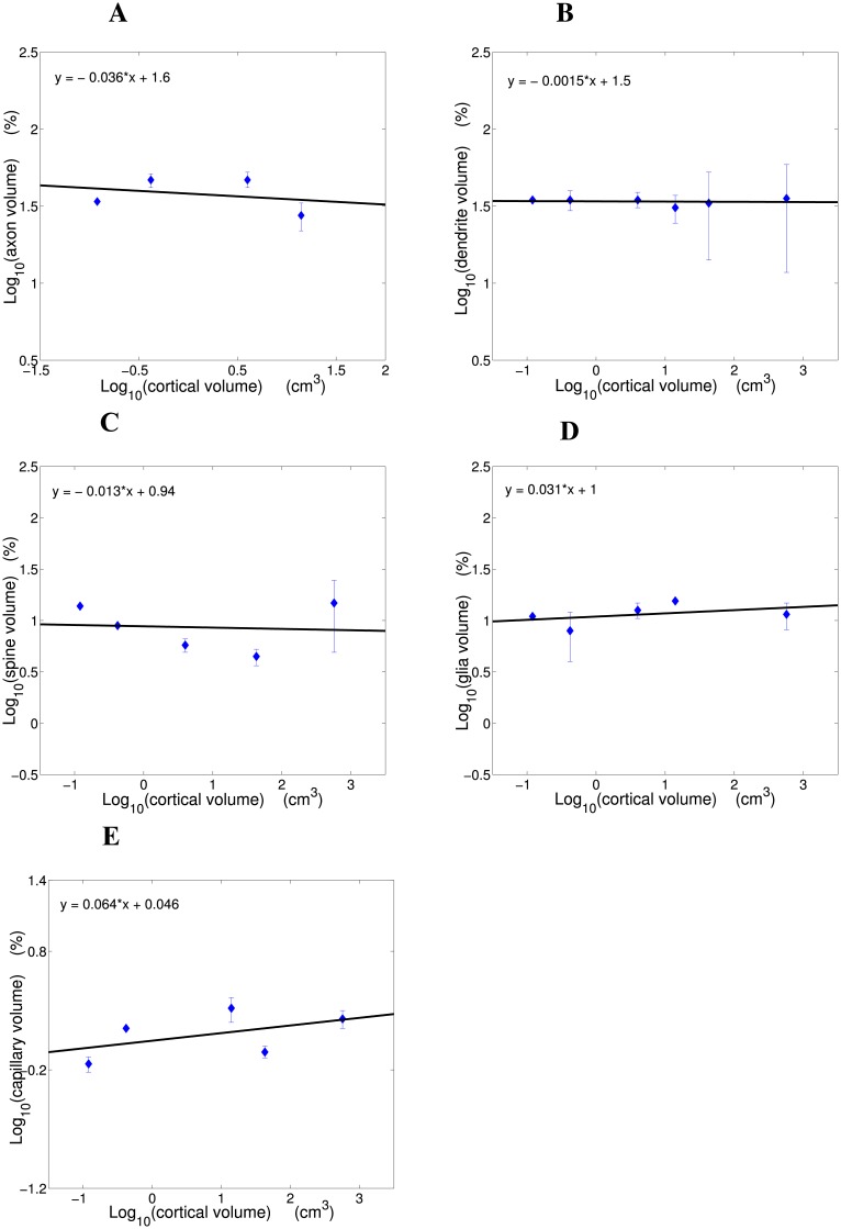
Fig 2. Scaling dependence of fractional volumes of the basic cortical components on cortical gray matter volume.
(A) Axon fractional volume, (B) dendrite fractional volume, (C) spine fractional volume, (D) glia fractional volume, and (E) capillary fractional volume as functions of cortical volume in log-log coordinates. Note a conservation trend across mammals. Scaling plots were constructed based on data in Table 1. The following volumes of cortical gray matter (two hemispheres) were used: mouse 0.12 cm3 [38], rat 0.42 cm3 [39], rabbit 4.0 cm3 [40], cat 14.0 cm3 [40], macaque monkey 42.9 cm3 [39], human 571.8 cm3 [39].

