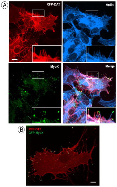
Figure 1. Membrane projection containing DAT are filopodia.
(A) PAE/RFP-HA-DAT cells were fixed, stained with phalloidin-Alexa680, permeabilized and immunostained with rabbit polyclonal MyoX antibodies followed by secondary antibody conjugated to Alexa488. 3D images were acquired through 488 (green, MyoX), 561 (red, DAT-RFP) and 640 (cyan, actin) channels. Maximal projection of the z-stack is presented. Insets show high magnification of the region marked by white rectangles.
(B) GFP-MyoX was transiently expressed in PAE/RFP-HA-DAT cells, and 3D live-cell imaging was performed through 488 (green, GFP) and 561 (red, RFP) channels as described in “Methods”. A single x–y confocal image of merged GFP and RFP fluorescence is presented.
Scale bars, 10 μm.

