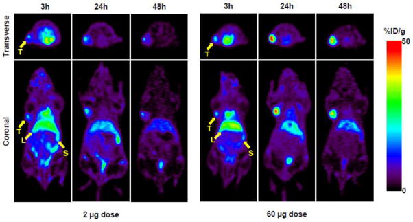
Figure 3.

Representative transverse and coronal PET images of 64Cu-NOTA-amatuximab (0.3 mCi) coinjected with 2 μg and 60 μg amatuximab at 3, 24, and 48 h p.i. in nude mice with tumor size ~200 mm3 (range, 170 to 320 mm3). PET images demonstrate that tumor uptake significantly increased when injection dose was 60 μg. T, tumor; L, liver; S, spleen.
